Grand Journal of Urology
E-ISSN : 2757-7163

Current Issue
Editorial
Dear Colleagues, Our journal titled Grand Journal of Urology (Grand J Urol), whose foundation studies were completed by the end of 2020 was published in January 2021 by publishing its first issue and took its place among scientific journals in the field of urology. The journal aims to publish original scientific urological articles. It is an open access, peer-reviewed journal and will be published online three times a year (January, May and September) in English. Our primary goal is to carefully evaluate the works of domestic and foreign authors, to take place in national and international reputable indexes with original and scientific articles, and to announce its name and content on scientific platforms. One of the most important criteria in the long journey to an academic career in our country is the production of scientific articles. I believe that the recently applied associate professorship criteria and the subsequent professorship criteria will increase the production of scientific articles in our country. This increase will increase the demand for distinguished, rigorous and scientific journals. In this process, the Grand Journal of Urology (GJU) will play an important role in delivering written and visual scientific publications to academic platforms and contributing to urology. GJU will add a new impetus to academic activities with its unique style. I am honored to present you the first issue of the Grand Journal of Urology (GJU) journal with the contributions of valuable researchers and scientists in a period when branch activities have decreased considerably due to the COVID-19 pandemic. At the beginning of this journey of our journal, I would like to express my wholehearted gratitude to the very valuable members of the Urology Community, my colleagues, my friends and my dear wife, who always supported us, our authors who contribute to our journal with orginal articles, review articles, case reports, clinical images and letters to the editor, our reviewers who meticulously evaluate the articles and present their support, our designers, and our publisher. January 2021 Editor-in-Chief Assoc. Prof. Ekrem Guner
Original Article
Andrology (Male Sexual Disfunction, Infertility), Online First: 14 December 2020
Grand J Urol 2021;1(1):1-5, DOI: 10.5222/GJU.2021.87597
Objective: Today, infertility is a health problem with increasing treatment seeking. Testicular sperm extraction (TESE) is the only possible procedure to offer genetic parenting to men with nonobstructive azoospermia (NOA). Our aim in this study is to present our clinical experiences that affect the success of sperm retrieval in men with NOA in the light of the literature. Materials and Methods: In our study, patients who underwent TESE with a diagnosis of NOA between 2017-2020 were retrospectively analyzed. According to the TESE procedure; the patients were divided into two groups as conventional TESE and TESE performed under microscopic magnification (micro-TESE). Medical histories, hormone values, and physical examination findings of all patients were recorded. Results: Our micro-TESE success rate was found to be 100%. A positive correlation (rho 0.714, p = 0.009) was found between the factors affecting sperm retrieval , and the application of micro-TESE, and a negative correlation was detected with FSH levels (rho -0.759, p = 0.004). Conclusion: The success of sperm retrieval increases with the micro-TESE procedure. As FSH levels increase, sperm retrieval success rates decrease.
Andrology (Male Sexual Disfunction, Infertility), Online First: 17 December 2020
Grand J Urol 2021;1(1):6-8, DOI: 10.5222/GJU.2021.43531
Objective: Varicocele is the abnormal venous dilatation and the tortuosity of the pampiniform plexus. Varicocele has been shown to be related with systemic varicosity in some studies. Platelet volume indices have also been reported to increase in vascular disorders. In this study, we aimed to determine if complete blood count (CBC) parameters especially platelet counts and volume indices could be a practical tool in the diagnosis and follow-up of varicocele. Materials and Methods: The medical records of all patients who underwent varicocelectomy due to grade 2 or 3 clinical varicocele were reviewed. Examined parameters included patient demographic characteristics and preoperative CBC parameters [hemoglobin, white blood cell, platelet, mean platelet volume (MPV) and platelet distribution width (PDW)]. Patients without varicocele, active infection and vascular disorders constituted the control group. Results: The study population consisted of 61 patients with varicocele and 62 control subjects. The mean age of the patients was 28.6 ± 6.2 years. Mean preoperative hemoglobin, WBC, platelet, MPV and PDW were 15.5 ± 1 g/dL, 7.5±1.6 x10³/μL, (236 ± 53.4) x103/μL, 9.3±1.1 (fL) and 15.2± 3.9 (%), respectively. There was no difference between patients with varicocele and control subjects in terms of age, mean preoperative Hb, WBC and MPV. However, mean preoperative platelet count was significantly lower and mean PDW was significantly higher in varicocele patients compared to controls (p
Andrology (Male Sexual Disfunction, Infertility), Online First: 22 December 2020
Grand J Urol 2021;1(1):9-13, DOI: 10.5222/GJU.2021.97269
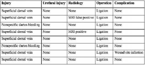
Objective: Penile fracture is one of the urological emergencies that require early surgical intervention. False penile fracture, on the other hand, is a condition that presents with similar clinical features and can be treated conservatively. In this study, in the light of the literature, it was aimed to present the clinical and operative results of 8 patients who were operated on with a prediagnosis of penile fracture and then diagnosed with a false penile fracture. Material and Methods: Data of 8 patients who were diagnosed with a false penile fracture between January 2006 and September 2019 were retrospectively analyzed. Patients" demographic characteristics, preoperative, intraoperative and postoperative data were retrospectively analyzed. Results: Mean age of the patients was 39.12 (28-54) years. The most common complaints were penile swelling and ecchymosis. The most common etiological factors were as follows: sexual intercourse in 6, masturbation in 1, and manual bending of the erect penis in 1 patient. All operations were performed by degloving the penis from the circumcision line. Superficial dorsal vein injury was detected in 6, and nonspecific dartos bleeding was detected in 2 patients. There were no intraoperative complications. Wound site infection developed in 1 patient postoperatively. No erectile dysfunction, penile curvature, and sensory disturbances were detected in any patient. Conclusion: It is difficult to distinguish a false penile fracture from true penile fracture clinically or radiologically. False penile fracture can be treated conservatively without the need for surgery. Surgery should still be the first-line treatment option in suspected patients. Studies with larger patient series are needed on this subject.
Functional Urology, Online First: 28 December 2020
Grand J Urol 2021;1(1):14-17, DOI: 10.5222/GJU.2021.47955
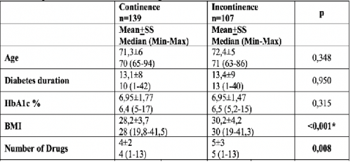
Objective: In our study, we aimed to examine the effects of body mass index (BMI) and multiple drug use on urinary incontinence (UI) in diabetic geriatric patients. Materials and Methods: Our study included 246 type 2 diabetes mellitus patients aged 65 and over who applied to our outpatient clinic between October and December 2019 and remained after the exclusion criteria were applied. The relationship between the frequency of UI and age, diabetes age, HbA1c, BMI, number of drugs and gender was investigated. UI described as any involuntary incontinence complaint. The questionnaire contained socio-demographic questions and the International Consultation on Incontinence Modular Questionnaire Urinary Incontinence Short Form (ICIQ-UI SF).with UI were women. Diabetes age and HbA1c level did not differ significantly between those with and without UI. There was a statistically significant relationship between BMI and the number of drugs used with the indication of UI (Mann-Whitney U; p
Endourology, Online First: 05 January 2021
Grand J Urol 2021;1(1):18-21, DOI: 10.5222/GJU.2021.47966
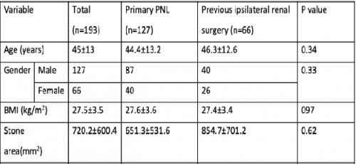
Objective: We aimed to compare outcomes of percutaneous nephrolithotomy (PNL) surgery in patients who had and had not undergone renal stone surgery before PNL. Material and Methods: We retrospectively analyzed the medical records of all patients who underwent PNL in our department between 2010 and 2019. Examined parameters consisted of patient demographics, medical and surgical history, stone size, stone density, stone site, estimated intraoperative blood loss, duration of operation, hospital stay and stone-free status. Results: A total of 193 patients were included the study. The mean age of the patients was 45±13 years. The mean duration of surgery was 69±11.5 minutes. The mean stone area was 720.2±600.4 mm2 and the mean stone attenuation was 982.8±327.7 HU. The mean postoperative hemoglobin decrease was 1.8±1.3 g/dL. 66 patients had previous stone surgery including open stone surgery, PNL and retrograde intrarenal surgery (RIRS). There was no statistically significant difference between patients who had and had not previousşy undergone renal stone surgery in terms of age, gender, body mass index and stone area. Operative time, estimated intraoperative blood loss, postoperative hemoglobin decreases and hospital stay were comparable between patients who had, and hed not undergone previous renal stone surgery. Stone-free rate was significantly higher in primary PNL patients compared to patients with a history of renal stone surgery (92.1% vs 77.3%, p=0.006). Conclusion: PNL has a similar complication rate in patients with and without previous kidney stone surgery. However, achieving stone-free status may be challenging in patients with a history of ipsilateral renal stone surgery.
Review Article
Urological Oncology, Online First: 22 December 2020
Grand J Urol 2021;1(1):22-25, DOI: 10.5222/GJU.2021.54264
Prostate cancer is one of the most common tumor in males. Radical prostatectomy, radiotherapy and watchful waiting are the main treatment options in localized disease. Radiotherapy together with hormonotherapy is accepted as the standard of care in patients with advanced stages. Surgery or radiotherapy has comparable local control and survival outcomes in localized disease. During recent years a significant reduction in the rate of serious side effects has been achieved due to the development of modern radiotherapy techniques. With the use of these techniques such as Intensity-modulated radiotherapy (IMRT), Image-guided radiotherapy (IGRT), Stereotactic body radiotherapy (SBRT), high doses can be given safely and the rates of serious short-or long-term side effects have not exceeded 1 percent. Modern radiotherapy techniques allow dose escalation for the target volume, and due to its achievement of sharp dose gradient around the target volume and enable to increase radiation doses homogeneously within the target volume without exceeding the tolerance doses in organs at risk. In the last few years hypofractionation has gained popularity in the curative radiotherapy of prostate cancer.
Functional Urology, Online First: 05 January 2021
Grand J Urol 2021;1(1):26-29, DOI: 10.5222/GJU.2021.57966
Overactive bladder is a serious condition that can significantly impair quality of life. Antimuscarinic agents are recommended as second-line therapy in patients who do not benefit from behavioral therapy. However, the therapeutic efficacy of antimuscarinic agents is limited. Alternative treatment methods to medical treatment have been developed due to its limited effectiveness and frequent side effects. Posterior tibial nerve stimulation (PTNS), transcutaneous tibial nerve stimulation (TTNS), sacral neuromodulation (SNM), intravesical Botulinum toxin-A (BoNT/A) are prominent among these minimally invasive treatment methods in refractory OAB. All these methods have been demonstrated to be effective in the literature. BoNT/A and SNM are more effective, but have been reported to cause more side effects. In refractory OAB, any of these methods can be applied by considering the medical condition and request of the patient.
Case Report
Female Urology, Online First: 29 December 2020
Grand J Urol 2021;1(1):30-32, DOI: 10.5222/GJU.2021.65375
Acute urinary retention is one of the most common situations encountered in urological emergencies. It is more frequently seen in older men. Acute urinary retention in pregnant women is a rare condition but it may cause abortus, preterm labor and rarely uterine ischemia. It is very difficult to reveal the causes of acute urinary retention in pregnant women. One of them is acute urinary retention due to retroverted uterus which is observed in 11% of pregnant women. In this case, we discussed a young pregnant patient who presented to our outpatient clinic with complaints of recurrent urinary retention due to retroverted uterus.
Urological Oncology, Online First: 06 January 2021
Grand J Urol 2021;1(1):33-36, DOI: 10.5222/GJU.2021.98609
Retroperitoneal liposarcoma (RPLS) is a rare tumor. Early diagnosis and treatment are difficult due to absence of specific clinical presentations. We report a case of a 66-years-old woman who succesfully underwent complete surgical resection for a giant retroperitoneal liposarcoma. The complete surgical resection is the most important predictor of local recurrence and overall survival. We believe that complete surgical resection involving adjacent organs is a curative treatment to increase overall survival, especially in the presence of invasion of large tumors.
Clinical Image
Genitourinary Radiology, Online First: 04 January 2021
Grand J Urol 2021;1(1):37-38, DOI: 10.5222/GJU.2021.66376
A 36-year-old male patient was admitted to the emergency department with abdominal and left side pain 1.5 hours after an in-vehicle traffic accident. The patient had no history of comorbidity or surgery. The patient did not receive any anticoagulant or antiplatelet therapy prior to the trauma. Vital signs of the patient were stable (Blood pressure 145/100 mmHg, pulse 98 beats/min, and temperature 37.2°C). Physical examination revealed no additional pathology except left side pain and left upper quadrant tenderness in deep palpation. White blood cells were found to be 17.330/mm3, hemoglobin level 17.34 mg/dL, and hematocrit 48.83% in the complete blood count examination. There was no abnormality in the biochemical analysis. An evaluation focused on sonography for trauma (FAST) in the emergency department was negative. Contrast-enhanced thoracoabdominal computed tomography (CT) scan was performed. Abdominal CT revealed a 44x42 mm left central hyperdense and peripheral hypodense adrenal mass (63 hounsfield unit) and periadrenal fat strands. Additionally, a cortical cyst was observed in the upper pole of the left kidney (Figure 1). The lesion was evaluated as an adrenal hematoma. No other injuries were detected, especially no injury to the spleen or kidney. No rib or spine fractures were observed. The patient was treated conservatively with bed rest, parenteral fluid, antibiotherapy, and analgesics. Hemoglobin and biochemical parameters remained constant. Endocrinology consultation was requested for adrenal insufficiency. Endocrinological evaluations revealed no pathology. The control abdominal CT performed 7 days later showed that the hematoma did not progress (40x37 mm, central density is prominent) (Figure 2). The patient was discharged without any problems. An abdominal CT scan was planned to evaluate the resolution of the adrenal hematoma 1 month later. The patient was asymptomatic during the follow-up. No abscess or infection format was observed. Informed written consent was obtained from the patient for this report. Figure 1: A- Left adrenal hematoma 44x42 mm attenuated central hyperdense and peripheral hypodense round mass in the adrenal gland, B- Periadrenal fat stranding, C- Left kidney upper pole simple cortical cyst (Axial view of enhanced abdominal CT scan) Figure 2: Control abdominal CT; regression of hematoma and increased appearance of central hyperdensity (Axial view of enhanced abdominal CT scan) Adrenal gland injury is a rare clinical picture caused by motor vehicle accidents, sports injuries, or blunt abdominal trauma after falling [–]. Isolated adrenal gland injury is rare due to its small size, deep retroperitoneal position on the upper abdomen, and presence of full-fat tissue around it. Most adrenal gland injuries are associated with multiple adjacent skeletal and organ injuries []. Adrenal gland injuries have been reported in approximately 2-3% of all thoracoabdominal injuries []. Unilateral adrenal injuries occur 5 times more on the right side than on the left (77% versus 15%), and bilateral adrenal injuries occur in 8% of cases in trauma []. Being usually silent and self-limiting, it does not require major operative intervention. However, it may be potentially life-threatening in some cases. The most common symptom is pain; other clinical presentations vary greatly, and it does not produce any specific symptoms or biomarkers. Abdominal pain, side pain, nausea, vomiting, hypotension, hypertension, a palpable side mass, agitation, mental status changes, and lowgrade fever may occur []. The emergency physician should be aware of the possibility of organ damage associated with adrenal injury and the potential for adrenal insufficiency especially if an unusual complaint is presented after blunt trauma (unexplained hypotension, electrolyte disorder, and pain that does not go away despite analgesics) []. Although ultrasonography is noninvasive, easily accessible, and inexpensive, it is dependent on the person and can sometimes be inadequate when evaluating retroperitoneal organs. CT is the gold standard for detecting adrenal gland injury as in all trauma cases []. CT scan findings of adrenal gland injury include hyperdensity, periadrenal fat stranding infiltration, and ipsilateral diaphragmatic crural thickening [,]. Furthermore, the need to monitor and rule out an underlying adrenal neoplasm should be taken into account in these patients due to possible bleeding to a pre-existing adrenal mass []. Surgery (adrenalectomy) and interventional radiologic procedures (embolization) may be needed although most adrenal gland injuries are treated conservatively. Treatment depends on the hemodynamic condition of the patient, the severity of the gland damage, bilateral gland involvement, and the extent of bleeding within the gland []. Ethics Committee Approval: N / A. Informed Consent: An informed consent was obtained from the patient. Publication: The results of the study were not published in full or in part in form of abstracts. Peer-review: Externally peer-reviewed. Conflict of Interest: The authors declare that they have no conflict of interest. Financial Disclosure: The authors declare that this study received no financial support.
Andrology (Male Sexual Disfunction, Infertility), Online First: 06 January 2021
Grand J Urol 2021;1(1):39-40, DOI: 10.5222/GJU.2021.91885
A 74-year-old male patient was admitted to the emergency department reaching a depth of 1 cm surrounding the penis body, bleeding, and discoloration of the penis skin. It was observed that there were white-yellow rubber bands in the incision area in the examination of the patient (Figure 1). Laboratory examinations revealed no pathology. The patient was consulted at the urology clinic. It was learned that he underwent urethral surgery after trauma and he had continuous urinary incontinence and compressed his penis with these rubber bands to prevent it. 18 Fr urethral Foley catheter was inserted. It was observed in the exploration that the rubber bands lasered the penis skin laterally and dorsally to tunica albuginea, and ventrally to corpus spongiosum and urethra level. Five rubber bands were cut and removed (Figure 2). It was observed that corpus spongiosum-urethra and corpus cavernosa were intact in exploration. The penile skin was left for secondary healing after sterile cleansing of the skin and subcutaneous tissue (Figure 3). Penis was wrapped with a Coban bandage after the medical dressing. The urethral catheter was removed on the first day after the operation. The patient was prescribed broad-spectrum antibiotherapy, analgesic, anti-inflammatory, and duloxetine for continence. Kegel exercises were practically explained. The patient was referred to the psychiatric clinic before discharge. It was observed in the follow-up one week later that the penis healing was good except for mild edema and the wound healed completely (Figure 4). The penis was found to be completely normal except for skin pigment change in several areas a month later (Figure 5). The patient stated that there was intermittent continence. Written informed consent form was obtained from the patient. Figure 1: Preoperative appearance Figure 2: Removed rubber bands Figure 3: Postoperative appearance Figure 4: Control appearance after 1 week Figure 5: Control appearance after 1month Penile strangulation with a foreign material is a rare condition and was first reported by Gauthier in 1755. To date, only a few case series have been published in the literature with fewer than 100 case reports. Penile strangulation is a condition that needs to be intervened urgently, and it can lead to complications such as gangrene and amputation of the penis if not treated as soon as possible [,]. Foreign materials used for strangulation can be classified as soft and hard. In the literature, the most common hard materials for strangulation were metallic rings (49.0%), metallic tubes (14.8%), plastic bottles (12.1%), rings (9.4%) and plastic products (6.7%) and the most common soft materials for strangulation were rubber bands (67.9%), rubber strings (13.2%), threads (13.2%) and vinyl products (1.9%). The most common causes to use foreign material for penile strangulation were pranks, sexual intercourses, treatments of incontinence, and treatments of phimosis []. Complications related to penile strangulation injuries are skin erosion, laceration, infection, urethral transection, penile gangrene, and autoamputation []. Bhat et al. developed a grading system for penile strangulation injuries due to constructive objects around the penis and divided them into five categories from penis edema to gangrene. Grade I causes edema only, whereas Grade II involves penile paresthesia. Grade III includes skin and urethral damage but does not include urethral fistula. Grade IV includes the urethral fistula. It involves Grade V injury, gangrene, necrosis, or complete amputation []. The management of the patients is different according to the type of foreign body and the clinical findings of each case. There is not a standard surgical approach []. The treatment mainly aims to remove the constricting object as soon as possible to restore venous and lymphatic drainage and arterial flow by preserving the anatomy and functionality of the organ []. Thin non-metallic constricting objects are easy to remove in the treatment of penile strangulation. Successful results can be obtained by cutting such objects with simple surgical scissors or a scalpel. Orthopedic surgical instruments or non-medical instruments may be needed in metal objects or in patients with severe edema after penile strangulation []. In addition, psychological and psychosexual evaluation of these patients is a part of the treatment. Ethics Committee Approval: N / A. Informed Consent: An informed consent was obtained from the patient. Publication: The results of the study were not published in full or in part in form of abstracts. Peer-review: Externally peer-reviewed. Conflict of Interest: The authors declare that they have no conflict of interest. Financial Disclosure: The authors declare that this study received no financial support.
Letter to the Editor
General Urology, Online First: 08 January 2021
Grand J Urol 2021;1(1):41-42, DOI: 10.5222/GJU.2021.98608
The new coronaviruses outbreak caused by SARS-CoV-2 (COVID-19) originated from the Chinese region of Wuhan in the last quarter of 2019 affected approximately 75 million people all around the world and caused over 1.6 million deaths []. COVID-19 is a highly contagious viral infection and its main routes for transmission are the person to person contact, touch, and aerosol. While it has detrimental effects on respiratory and cardiovascular systems it also can be found in digestive and urinary systems. The frequently experienced symptoms are fever, dry cough, dyspnea, fatigue, and loss of appetite []. With an ongoing effort, several researchers focused on investigating a drug or vaccine to end the pandemic. Currently, despite there is no drug specifically approved for COVID-19 treatment, there has been more than one vaccine from different nations to prevent the virus spread. Prior to the COVID-19 pandemic, virology research constituted less than 2% of all biomedical research. But this rate has been increased to 10-20% which represents the incredible adaptation potential of the research community. By the way, the COVID-19 pandemic led to a massive influx of publication not only by virologists and infectious health specialists but also by almost all medical disciplines. To facilitate early dissemination of knowledge prior to any peer-review, many articles have been uploaded preprint services []. It is not realistic to assume that a qualified and strict peer-review process could compensate for the high number of submissions. Moreover, someone must be aware that those non-peer-reviewed materials could be picked up by the media and spread to the population. Social distancing and transmission issues have also led to travel and social restrictions that resulted in many trials to be suspended or delaying in patient recruitment []. It is wellknown that generally large-scale randomized trials were not set up in time in the previous pandemic. However, the use of modern information technologies in combination with oldfashioned randomization might lead to the rapid gain of viable results nowadays. Countries might be encouraged to establish clinical-trial networks to activate and arrange large multi-center studies []. COVID-19 pandemic deeply affects not only urological patient care but also urology residency education. Work hours modified and residences redeployed to serve in pandemic services in many countries. This situation came along with the problem of interrupted urologic training and unmet minimal case requirements. Generally, online learning curricula have been well-adopted by urologists. However, recent studies have been shown that such changes caused emotionally and physically stressful situations for trainees []. In conclusion, it is obvious that Coronavirus will continue to be in our daily life for a while. Thus, maintaining research and education is vital in all disciplines of medicine. Contributing to the scientific area with respect to essential requirements and ethics will support the development of all humanity in this crisis. Hereby, We would like to congratulate you on the first issue of your journal planned to be released in such a difficult time and wish you to have significant contributions to the field of urology in a strictly scientific manner. Sincerely yours.