Grand Journal of Urology
E-ISSN : 2757-7163

Current Issue
Editorial
Editorial, Online First: 20 September 2024

Dear colleagues, I am honored to share with you the second issue of 2024 (volume 4, issue 3) of the Grand Journal of Urology (Grand J Urol) with the contributions of many respected researchers and authors. Grand Journal of Urology (GJU) aims to carry written and visualscientific urology studies to academic platforms and to make significant contributions to the science of urology. Our journal has been abstracted/indexed in Tubitak Ulakbim TR Index, EBSCOhost, J-Gate, SciLit, ResearchGate and Google Scholar international databases. As of these achievements, the Grand Journal of Urology (GJU) has taken its place among the journals indexed by national and international databases. In this issue of our journal, there are many valuable articles under the subheadings of Functional Urology, General Urology, Urological Oncology and Urolithiasis. I hope that these carefully prepared articles will make important contributions to valuable readers, researchers and the urology literature. On this occasion, I would like to express my heartfelt gratitude to our authors who have contributed to our journal with their articles, to our reviewers who have meticulously evaluate the articles. Respectfully yours September 2024 Assoc. Prof. Ekrem GUNER, MD Editor-in-Chief
Original Article
Urological Oncology, Online First: 09 August 2024
Grand J Urol 2024;4(3):070-076, DOI: 10.5505/GJU.2024.18894
Objective: To evaluate whether the assessment of anxiety and pain perception before a biopsy procedure may predict patients" perceived pain scale scores during transrectal ultrasound-guided prostate biopsy. Materials and Methods: Patients who were administered the Mini-Mental State Examination 24 h before the biopsy were evaluated based on electrically and mechanically induced pain thresholds. Patients were assessed with Generalized Anxiety Disorder (GAD)-7 scale sscores in the hour before biopsy. The pain experienced by patients during rectal probing and biopsy was assessed using Visual Analog Scale (VAS) scores. Results: The mean age and median PSA level of the patients were 65.52 ± 7.85 years and 9.73 (1.4-155) ng/dL, respectively. The median VAS scores during rectal probing and biopsy were 3 (0-10) and 4 (0-10) respectively. VAS scores calculated during procedures were moderately-to-strongly correlated with the index finger of mechanically induced pain pressure threshold (PPT) (r=−0.606, p=0.001 and r=−0.760, p=0.001). Multiple regression analyses revealed that severity of the intraprocedural pain was correlated with age, GAD-7, and PPT index finger scores (p=0.005, p=0.001, p=0.001, respectively). A correlation was noted between the refusal of repeat prostate biopsy and higher pain scores (p
Urolithiasis, Online First: 29 August 2024
Grand J Urol 2024;4(3):077-082, DOI: 10.5505/GJU.2024.30092
Objective: To identify patient- and procedure-related factors that increase the risk of hospital readmission (HR (and emergency room (ER) visits after percutaneous nephrolithotomy (PNL). Materials and Methods: Patients who underwent supine PCNL operation between 2018 and 2023 were retrospectively reviewed. Patient demographic characteristics including age, body mass index, ASA score, stone size, presence of anatomical abnormalities and comorbidities, preoperative and postoperative data, and emergency department visit and readmission rates were analysed. ER to HR, including elective ones, and ER visits for any reason related to the PNL procedure were primarily analysed. Factors affecting the rate of ER visit and HR were analysed using logistic regression analysis. Results: The mean age of 450 patients who underwent supine PCNL was 42.1 ± 20.8 years. When SFR was accepted in < 4 mm fragments, the SFR rate was 87%. Complications were observed in 19.5% of patients. ER rate was 8.8% and HR rate was 7.7%. Anatomical abnormality, stone complexity, operation time and postoperative complications were statistically significant for ER, while comorbidity, high ASA score, anomalous kidney, stone complexity, long operation time and postoperative complications were statistically significant for HR. Conclusion: In our study, unplanned hospitalization was observed at a higher rate in patients with anatomical abnormalities and complex kidney stones. HR and ER were found more frequently in patients with a history of complications.
Urological Oncology, Online First: 02 September 2024
Grand J Urol 2024;4(3):083-088, DOI: 10.5505/GJU.2024.54227
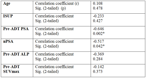
Objective: We sought to identify predictive factors affecting time to castration resistance in metastatic prostate cancer patients receiving androgen deprivation therapy (ADT). Materials and Methods: We retrospectively evaluated 47 patients who received ADT with the diagnosis of metastatic prostate cancer. The patients" age, International Society of Urological Pathology (ISUP) scores, baseline prostate specific antigen (PSA), alkaline phosphatase (ALP) value, prostate-specific membrane antigen (PSMA) tracer expression, represented by the maximum standardised uptake value (SUV max) at diagnosis, nadir PSA value, and time to resistance to ADT were recorded. Results: All patients included in the study were resistant to treatment with ADT. The mean age of the patients was 70.81 ± 1.15 years. The mean time to develop resistance to treatment after castration was 31.51 ± 4.9 months. In the correlation analysis, a significant negative correlation was detected between PSA, nadir PSA values and time to treatment resistance. The relationship between the SUVmax value of the primary prostate lesion, ALP value at the time of diagnosis and time to response to ADT developed was not significant. Conclusion: We found PSA values at diagnosis and nadir PSA values during follow-up to be predictive factors of treatment resistance in metastatic prostate cancer patients receiving ADT.
Functional Urology, Online First: 06 September 2024
Grand J Urol 2024;4(3):089-097, DOI: 10.5505/GJU.2024.77699
Objective: To evaluate the pain characteristics of women with overactive bladder (OAB) for investigating the role of central sensitization in OAB pathophysiology. Materials and Methods: Women with OAB over the age of 18 years and healthy volunteers made up the participants in the current study. Pain intensity and quality were analysed with the Short Form of the McGill Pain Questionnaire (SF-MPQ). The Self-Leeds Assessment of Neuropathic Symptoms and Signs (LANSS) was used to assess the presence of neuropathic pain. Pain threshold was evaluated with algometer. The Pressure Pain Threshold measurement was determined as the primary outcome measure of the present study. The Overactive Bladder Awareness Tool (OAB-V8), short forms of the Incontinence Impact Questionnaire-7 (IIQ-7) and Urogenital Distress Inventory-6 (UDI-6) were used to evaluate OAB symptoms. Nottingham Health Profile (NHP) questionnaire was used to reveal quality of life and general health status. Results: According to algometric measurements, OAB patients had lower pain thresholds in 19 anatomical points (p
Case Report
Urological Oncology, Online First: 10 June 2024
Grand J Urol 2024;4(3):098-102, DOI: 10.5505/GJU.2024.52714
Six percent of cases with renal cell carcinoma (RCC) can present with thrombus, and also invasion to renal vein, and atrium may be observed in 44% and 1-4 % of these cases, respectively. These cases require multidisciplinary management and surgery should be the first treatment option. However, if a tumor is considered unresectable or metastatic, systemic therapy can be considered in the first instance. Herein, we present 2 cases. A 77-year-old female patient presented with right renal tumor 89 mm in diameter with thrombus level IV considerably unresectable started to receive treatment with nivolumab and cabozantinib. After 6 months of treatment thrombus was reduced to level II. A 43-year-old male, presented with 110 mm- right renal mass with thrombus level II and lung metastases. He started to receive pembrolizumab and axitinib. At 6 months of treatment, the size of the tumor and thrombus decreased. In both cases we performed laparoscopic radical nephrectomy with thrombectomy, and pathology reports indicated the presence of clear cell RCC, Grade 3, pT3b-Nx. Systemic treatment in patients with RCC associated with tumor thrombus, whether metastatic or not, would seem to obtain some benefit prior to surgery -line favor surgical feasibility.
Urological Oncology, Online First: 13 August 2024
Grand J Urol 2024;4(3):103-105, DOI: 10.5505/GJU.2024.53386
Extra-uterine leiomyomas of urethral origin are rarely encountered neoplasms possessing unique features such as the characteristic growth pattern, diagnostic challenges owing to a long list of possible differential diagnoses, possible cure with surgical management and the unique complications that accompany surgical management. Herein, we report a case of urethral leiomyoma in a middle- aged woman with a brief discussion on the evaluation and management aspects including a concise description of this pathology based on the scarce literature information available.
Clinical Image
General Urology, Online First: 14 August 2024
Grand J Urol 2024;4(3):106-107, DOI: 10.5505/GJU.2024.07379
A 22-year-old woman presented to the emergency department with a 2-hour history of abdominal/flank pain. She was involved in a motor vehicle collision where she was the driver. Airbags were deployed, but her seat belt compliance was unknown at the time of injury. Her medical history was mysterious and unattainable due to her altered mental status. On general appearance, the patient appeared intoxicated. Physical examination was only significant for abdominal tenderness to palpation. Vital signs revealed hypotension (97/64 mmHg). All other values, such as pulse, temperature, oxygen saturation, and respiration, were within normal limits. Laboratory values on admission revealed elevated transaminases (AST 117/ ALT 86), and urinalysis showed hematuria (RBCs >182/HPF). All other values were within the normal range. A FAST (focused assessment with sonography in trauma) ultrasound was subsequently done, which revealed free fluid collection within the abdomen. A multidisciplinary team, including urological surgery, was consulted. A subsequent computed tomography (CT) scan of the abdomen and pelvis showed intraperitoneal extravasation of contrast, consistent with dome rupture (Figure 1). Exploratory laparotomy was performed after and revealed a rupture across the bladder dome (Figure 2). The bladder was surgically repaired (3-0 Vicryl), and a Foley catheter was placed for twelve days. On follow-up, a cystogram was performed, which confirmed bladder healing, and the catheter was removed. The postoperative course was uncomplicated, and the patient completely recovered after two months. Figure 1. A computed tomography scan of the pelvis showing extravasation of contrast, which is consistent with an intraperitoneal bladder rupture. A: coronal view; B: sagittal view Figure 2. Laparoscopic view of the abdomen demonstrating free bloodstained fluid in the pelvic cavity and rupture across the dome of the bladder Bladder rupture is a rare condition due to the protection of the bladder by the sturdy pelvic bones []. Today, bladder injuries remain relatively uncommon, accounting for only up to 10% of abdominal trauma [-]. Although motor vehicle collision is the most common cause of injury, intragenic causes, including surgical and endoscopic procedures, have also been identified [,]. Bladder rupture can be divided into intraperitoneal or extraperitoneal rupture []. Extraperitoneal injuries are the most common among the two, accounting for approximately 80% of cases, with a general association of pelvic fracture with damage to the bladder trigone, neck, or wall []. Extraperitoneal injuries are commonly treated conservatively (with catheter drainage via foley or suprapubic tube) []. Most bladder ruptures, regardless of the classification, typically manifest with symptoms of pelvic pain with difficulty voiding and gross hematuria [-]. Intraperitoneal injuries, on the other hand, account for 15% of bladder injuries []. This typically occurs when there is a compressive force against a full bladder, which ruptures the weakest portion (dome) as presented in this patient [,]. A FAST ultrasound may be positive as urine accumulates in the abdominal cavity [,]. Treatment includes surgical repair, which has demonstrated high success rates [-]. Recent practical guidelines regarding intraperitoneal bladder injuries suggest surgical repair due to a more considerable risk for lacerations with poor wound healing, electrolyte derangement, and peritonitis [-]. According to the American Urological Association (AUA) guidelines, extraperitoneal injuries should be managed conservatively [-]. Acknowledgements: I would like to sincerely thank Dr. Matthew Meece (Department of General Surgery, University of Miami Leonard M. Miller School of Medicine/Jackson Memorial Hospital, Miami, FL, USA) for his assistance in interpretation of the images. Ethics Committee Approval: Not applicable, as this is an anonymous clinical image. Informed Consent: The patient in this study provided written informed consent prior to participation. Publication: The results of the study were not published in full or in part in form of abstracts. Peer-review: Externally peer-reviewed. Authorship Contributions: Any contribution was not made by any individual not listed as an author. Concept – K.E.; Design – K.E.; Supervision – K.E.; Resources – K.E.; Materials – K.E.; Data Collection and/or Processing – K.E.; Analysis and/or Interpretation – K.E.; Literature Search – K.E.; Writing Manuscript – K.E.; Critical Review – K.E. Financial Disclosure: This article received no specific grant or support from any public or private agencies.