Grand Journal of Urology
E-ISSN : 2757-7163

Urolithiasis
Original Article
Objective: This study aimed to investigate the effect of stone density on the success of ureterorenoscopy (URS) and retrograde intrarenal surgery (RIRS). Materials and Methods: The data of patients who underwent URS or RIRS due to kidney and ureteral stones between January 2013 and March 2018 were retrospectively screened. For all patients, age, gender, comorbidities, the American Society of Anesthesiologists (ASA) score, the presence of preoperative double-J (DJ) stents, extracorporeal shock wave lithotripsy (ESWL) history, ipsilateral stone surgery history, the presence of renal anomalies, stone laterality, stone opacity, stone density, stone size, stone volume, operative time, stone-free status, and the presence and size of residual stones were recorded. Results: The study included 566 patients who underwent URS or RIRS, including 186 women (32.9%) and 380 (67.1%) men. The mean age of the patients was 47 years. The mean stone size was 10 mm, and the mean stone density was 886 Hounsfield units. The mean stone volume was 426.13 mm3. The mean operative time was 31 minutes. The stone-free rate was 89.4%. Stone density, stone size, and stone volume were positively correlated with operative time (p
Grand J Urol 2023;3(2):042-048, DOI: 10.5505/GJU.2023.70883
Objective: We aimed to determine the individualized management of middle-sized kidney stones in the lower pole calyces that can be removed using shock wave lithotripsy (SWL) (Group A), flexible ureteroscopic retrograde intrarenal surgery (RIRS) (Group B) and micro-percutaneous lithotomy (micro-PNL) (Group C). Materials and Methods: Patients who had 1-2 cm kidney stones in the lower pole calyces whose calyceal necks (length: 5mm), pelvicalyceal angle (>30o) and relatively shorter stone-skin distance as determined based on tomographic urography results were included in the study. Patients with renal cystine, whewellite stones or stones with a hardness above 1000 Hounsfield units were excluded. The groups were not formed randomly. Contarily, treatment methods were explained to the patients and let them decide the treatment method for themselves. Each group consisted of 34 patients. Results: After excluding nine patients who were lost to follow-up, the study was completed with 93 patients at the final analysis. Stone-free rate was lower in Group A (47%) than Groups B (80.5%) and C (77%) (p
Objective: To investigate the effect of ureterorenoscopic stone removal timing on kidney function in unilateral ureteral stones. 
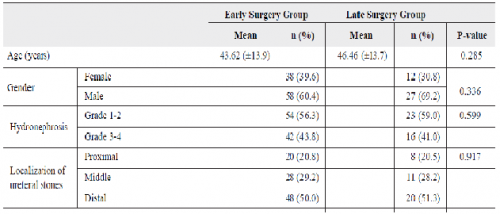
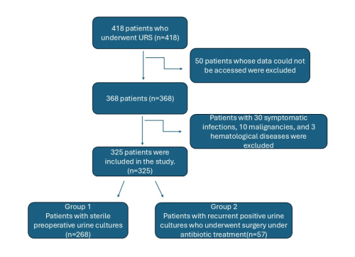
Materials and Methods: Hundred and eighty-seven patients included in the study were divided into two groups: 98 patients who underwent surgery ≤14 days after the stone diagnosis constituted the Early Surgery Group and 39 patients who were operated >14 days after the stone diagnosis comprised the Late Surgery Group. Preoperative serum levels of creatinine, blood urea nitrogen (BUN), and glomerular filtration rates (GFR) were recorded for the patients in both groups. In the postoperative first month, serum creatinine, BUN, and GFR were again recorded and compared with the preoperative values. 
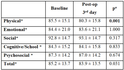
Results: The mean preoperative serum creatinine, GFR, and BUN levels in the Early Surgery Group were 1.25 ± 0.65μmol/L, 80.04 ± 33.6ml/min/1.73m2, and 50 ± 16.6mmol/L, respectively. A decrease was observed in serum creatinine (0.82 ± 0.22μmol/L, p< 0.001) and BUN (14.08 ± 7.25mmol/L, p< 0.001) levels one month after surgery, whereas GFR increased (105.33 ± 21.6ml/min/1.73m2, p< 0.001). In the Late Surgery Group, postoperative levels of serum creatinine (0.94 ± 0.33 vs. 0.95 ± 0.30μmol/L, p= 0.102), and BUN (17.38 ± 9 vs. 17.92 ± 8.8mmol/L, p= 0.283), increased minimally, also a minimal decrease was observed in GFR (95.15 ± 27.3 vs. 93.77 ± 24.3ml/min/1.73m2, p= 0.338) without any statistically significant difference. Conclusion: We believe that surgical treatment should be planned within two weeks at the latest, as prolonged obstruction may result in kidney damage.
Grand J Urol 2023;3(3):113-120, DOI: 10.5505/GJU.2023.63325
Objective: To compare extracorporeal shock wave lithotripsy (ESWL) induced renal injury in patients undergoing different ESWL treatment protocols by measuring urinary tissue metalloproteinase-2 inhibitor (TIMP-2) and insulin-like growth factor binding protein 7 (IGFBP7) excretion. Materials and Methods: This prospective, randomized study was conducted between April 2016 and June 2016 in group 1 patients undergoing fixed voltage ESWL and group 2 patients undergoing ramping voltage ESWL. Urinary TIMP-2 and IGFBP7 levels were analyzed before ESWL and 2 hours after ESWL, and urinary beta- 2-microglobulin (β2-MG) and albumin were analyzed before ESWL and 1 week after ESWL to assess renal injury. The primary outcome was to compare the effect of ESWL on early renal injury with biochemical markers in the different treatment protocols, and the secondary outcome was to compare the two treatment protocols in terms of stone free rate and complications. Results: There was no statistically significant difference between groups in terms of demographic and stone characteristics. There were statistically significant differences in serum creatinine and e-GFR at baseline and one week after treatment (p
Case Report
A hanging bladder calculus on the dome of the bladder is rarely seen, and a hanging bladder stone concurrent with bladder cancer is observed even rarer. Herein, we report a 76-year-old male patient presenting with lower urinary tract symptoms and recurrent urinary tract infection. A hanging stone on the dome of the bladder was seen and treated endoscopically. We also coincidentally found and resected a suspicious lesion which was diagnosed as low-grade papillary urothelial carcinoma.