Grand Journal of Urology
E-ISSN : 2757-7163

Current Issue
Editorial
Dear Colleagues, I am honored to share with you the second issue of the Grand Journal of Urology (Grand J Urol) with the contributions of valuable researchers and scientists. The journal is an open access, peer-reviewed journal and is published online in English three times a year (January, May and September). The Grand Journal of Urology (GJU) brings written and visual scientific urology studies to academic platforms and makes significant contributions to the science of urology. In addition, GJU adds a new momentum to academic activities with its unique style. After the first issue of our journal, valuable academicians from Autria, Germany, France, Italy and China joined the article review and advisory boards. In the meantime, I would proudly like to state that our journal has started to be indexed in the SCILIT database (an open-access scholarly database, Basel, Switzerland) and ScienceGate. In this second issue of our journal, there are many valuable articles under the subheadings of Genitourinary Radiology, Urological Oncology, General Urology, Andrology and Pediatric Urology. I hope these articles will provide important contributions to our dear readers and researchers. On this occasion, I would like to express my heartfelt gratitude to our authors who have contributed to our journal with their articles, to our reviewers who have meticulously evaluate the articles, to our designers and to our publisher. Respectfully yours May 2021 Assoc. Prof. Ekrem GUNER, MD Editor-in-Chief
Original Article
Urological Oncology, Online First: 05 April 2021
Grand J Urol 2021;1(2):43-48, DOI: 10.5222/GJU.2021.58066
Objective: Levels of insulin-like growth factor 1 (IGF-1) have been associated with prostate carcinoma. We have investigated whether IGF-1 level has an early predictive value for the biochemical relapse in the prostate carcinoma patients with a negative surgical margin who underwent curative surgery. Materials and Methods: We retrospectively analyzed 82 patients who were followed-up regularly and did not receive neoadjuvant or adjuvant chemotherapy. We classified patients as having Gleason scores ≥7 and
Andrology (Male Sexual Disfunction, Infertility), Online First: 22 April 2021
Grand J Urol 2021;1(2):49-54, DOI: 10.5222/GJU.2021.65376
Objective: The aim of this study is to investigate the effect of hyperlipidemia on the development of erectile dysfunction (ED) in hyperlipidemic patients with ED. Materials and Methods: Twenty-five patients who applied to the radiology clinic were included in the study. All patients have only hyperlipidemia as a risk factor of ED. The patients were evaluated in terms of ED by using International Index of Erectile Function (IIEF) form. Before and after oral treatment with daily doses of 10 mg atorvastatin, all parameters were measured. Paired t-test was used to compare vascular velocities between lipid profiles and Erectile Function Domain Scores (EFDS) and IIEFs, before and after treatment separately. Results: Cholesterol levels of 96% of patients were higher than 200 mg/dl and 52% of them had abnormal penile Doppler ultrasonography (PDU) findings. Patients with abnormal PDU findings had lower cholesterol levels than those with normal PDU findings. Significant differences existed between patients with normal and abnormal PDU in the high triglyceride group as for pre-, and post-treatment values . Pre-, and post-treatment EFD and IIEF scores were comparable. Conclusion: It can be said that a relationship exists between hyperlipidemia and erectile dysfunction. Therefore, lipid profile of a patient admitted with ED may be analyzed routinely
General Urology, Online First: 26 April 2021
Grand J Urol 2021;1(2):55-61, DOI: 10.5222/GJU.2021.36855
Objective: Complete urinalysis (CUA) is one of the indispensable screening tests of clinical laboratories. The compatibility of this test with urine culture is of indispensable importance in the diagnosis and treatment of urinary tract infections. We aimed to evaluate the suitability of the leukocyte parameter measured in the microscopic units of the fully automated urine analyzers which replace traditional methods, by grouping them according to the results of chemical analysis. Materials and Methods: Leukocyte counts in the reported CUA results of 4685 outpatients and the results of 113 urine cultures studied on the same day were analyzed. Noncentrifugated urine samples were included in the analysis. Cells were digitally imaged by flow microscopy. Chemical analyzes were performed using dual wavelength reflectance method. Urine samples were evaluated after 24 hours of incubation. 
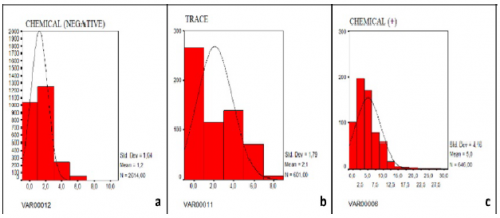
Results: High power field (HPF) values were recorded by grouping the leukocyte counts as negative, trace, 1+, 2+ and 3+. The arithmetic means of HPF values of the groups were calculated as 1.2, 2.1, 5.0, 11 and 208 white blood cell (WBCs/HPF). Bacterial growth was detected in 19 of 113 patients and no reproduction was observed in the remaining 94 cases. When results of microscopic examinations and chemical analysis were compared with the culture results, the analytical sensitivity, specificity, positive, and negative predictive values for microscopic urinalysis were 25%, 86%, 61.3%, and 58.3%, respectively. While, the analytical sensitivity, specificity, positive, and negative predictive values for chemical analysis of urine were 25.8%, 87.7%, 69.7%, and 51.4%, respectively. Conclusion: The workload of medical laboratories is increasing, and the use of urine autoanalyzers may be preferred for busy laboratories. In the diagnosis and follow-up of urinary tract infections, complete urinalysis by autoanalysers in which the harmony of their microscopy and chemical units are closely monitored, may reduce the need for unnecessary requests for urine culture, but it cannot replace urine culture.
Pediatric Urology, Online First: 11 May 2021
Grand J Urol 2021;1(2):62-65, DOI: 10.5222/GJU.2021.47956
Objective: Hypospadias is one of the most common congenital defects in boys. Multifactorial factors such as genetic predisposition and environmental factors play a role in the etiology of hypospadias. In this study, we investigated the risk factors of patients diagnosed with hypospadias. Materials and Methods: Thirty-six patients who applied to the pediatric endocrinology and urology outpatient clinics with the diagnosis of hypospadias were evaluated retrospectively. Risk factors were evaluated by recording the parental ages, exposure to environmental factors, the maternal BMI, history of pregnancy, drug use, and the father's fertility status. Results: The mean age of the patients was 3.5 ± 2 years. The patients had anterior (n:27 : 75%), middle (midshaft) (n:8 ; 22.2%), and posterior (n:1 ; 2.8%) hypospadias. The mean body mass index (BMI) of the mothers was 24 ± 4.1 kg/m2. Eight (22.2%) mothers were overweight and six (16.6%) mothers were obese. There was a history of hypospadias in the family of 4 (11%) patients. Conclusion: Although combinations of environmental and genetic factors play a role in the etiology of hypospadias, many unexplained factors are responsible for this disease.
Urological Oncology, Online First: 17 May 2021
Grand J Urol 2021;1(2):66-70, DOI: 10.5222/GJU.2021.39974
Objective: To investigate the value of D-dimer, a marker of fibrinolysis, in metastatic and non-metastatic prostate cancer. Materials and Methods: A retrospective analysis was performed on 138 male patients including 52 patients with prostate cancer and 86 with benign prostatic hyperplasia. Participants who had factors that altered D-dimer levels were excluded. The mean ages of the groups were similar (70 ± 8 vs 68 ± 8, p= NS). In addition, data regarding biochemical findings, prostate-specific antigen and hemostatic markers, including D-dimer, were retrieved from the database of our hospital. The cut-off point of D-dimer was 0.5 mg/L. Data from scintigraphy and magnetic resonance imaging (MRI) scans related to metastasis were also considered. Patients who showed findings of metastasis according to scintigraphy and lumbosacral MRI were accepted as having metastases. Positive findings in only scintigraphy in any area were considered suspicious for metastasis. Results: Patients with prostate cancer had higher D-dimer levels than benign prostate hyperplasia patients (p= 0.024). Sixteen patients with metastatic prostate cancer and suspicious for metastasis had markedly high D-dimer levels compared to benign prostate hyperplasia and non-metastatic prostate cancer patients. Conclusion: prostate cancer, especially when metastatic, may increase D-dimer levels.
Case Report
General Urology, Online First: 10 February 2021
Grand J Urol 2021;1(2):71-74, DOI: 10.5222/GJU.2021.25743
Hemorrhagic cystitis (HS) is a frequently seen complication of bone marrow transplantation. This condition occurs depending on the preparatory regimen of bone marrow transplantation. The BK virus (BKV), a human polyomavirus, is a small double helix DNA virüs belonging to the Papovaviridae family. It is commonly found in societies as an occult infection. Whereas, cidofovir (CDV), an acyclic nucleoside phosphonate, is used as a proven antiviral agent against polyomaviruses. In this case report, acute proximal tubular necrosis due to cidofovir used in hemorrhagic cystitis caused by BK virus, and its treatment in a patient diagnosed with T-lymphoblastic lymphoma in remission, and underwent allogeneic stem cell transplantation, was presented.
Andrology (Male Sexual Disfunction, Infertility), Online First: 10 February 2021
Grand J Urol 2021;1(2):75-77, DOI: 10.5222/GJU.2021.25745
Testicular torsion is a urological emergency that results in deterioration of the blood supply of the testicle and ischemia as a result of the rotation of the spermatic cord around itself. It may show a wide clinical variety with inflammatory manifestations varying from mild abdominal pain to severe scrotal pain. Orchiectomy may be required in cases which are delayed and cannot be operated urgently. Torsion of the testis and epididymis are other frequently seen causes of acute scrotum in children. Growth of masses and hormonal stimulation in the adolescent age cause an increase in the tendency of the torsion of appendix testis which have a small pedicle and epididymis. In the presence of sudden scrotal pain, testicular torsion should be considered, if there is clinical suspicion, patients should be evaluated with color doppler ultrasound (CDUS) and scrotal exploration should be performed immediately. A 20-year-old male whose clinical picture, and scrotal ultrasonography suggested the presence of testicular torsion is presented in this case report.
Urological Oncology, Online First: 02 April 2021
Grand J Urol 2021;1(2):78-80, DOI: 10.5222/GJU.2021.00710
COVID-19 is a highly contagious disease. This condition affects the decision of both the patient and the surgeon about the surgery of newly diagnosed cancer patients and it may also result in delays in cancer surgeries because of the limitations in healthcare applications. In our particular case, it was aimed to present the transition of the cancer from the localized stage to distant spread stage cancer since the patient who was pre-diagnosed with RCC and who was recommended surgery, did not want to undergo surgery due to COVID-19 pandemic and its risks. Our case was a 49-year-old female patient. In her computed tomography, a 58x70 mm heterogeneously enhancing solid lesion which showed exophytic extension from the middle zone of the right kidney to the lower pole was observed. Surgery was recommended for the patient but the patient claimed that she did not want to undergo surgery due to the risk of COVID-19 pandemic. The patient made an application for the surgery 8 months later. The new magnetic resonance imaging of the patient showed that there was a mass lesion of approximately 76x76x80 mm in size, which involved middle-lower part of the right kidney and extended into the opening of the renal vein VCI by invading the renal vein. Radical nephrectomy and thrombectomy procedure was applied to the patient with RCC? tumor. Due to psychosocial problems caused by the pandemic, surgeries are delayed and an acceleration of the cancer progression from the localized stage to the distant spread stage occurs indispensably.
Clinical Image
Genitourinary Radiology, Online First: 29 March 2021
Grand J Urol 2021;1(2):81-82, DOI: 10.5222/GJU.2021.09609
A 67-year-old male patient who had multiple solid masses in both kidneys in another center ultrasound examination was referred to our clinic for further examination. He had no known chronic or syndromic disease. The patient was evaluated with dynamic contrast enhanced upper abdominal magnetic resonance imaging (MRI). In MRI, homogeneous enhancing solid lesions with hypointense central stellate scar and non-enhancing cysts was seen in both kidneys. Diffusion restriction was observed in the periphery of the solid lesion in the lower pole of right kidney which was considered suspicious for malignancy and biopsy was performed from this lesion (Figure 1). Pathology result reported as oncocytoma so follow-up decision was made for the patient. In the follow-up, the masses in the left kidney increased in size and diffusion restriction developed in the upper pole of the left kidney (Figure 2). This lesion was also biopsied because of suspect imaging finding, the result was reported as oncocytoma as well. At the same time, the patient was evaluated for tuberous sclerosis and Birt-Hogg-Dube syndrome as they associated with multiple bilateral oncocytomas. There was no similar signs, semptoms and imaging findings related to these genetic syndromes in the family members of the patient. Informed written consent was obtained from the patient for this report. Figure 1: A-Homogeneous enhancing, B- Multipl solid lesions with hypointense central stellate scar and non-enhancing cysts in both kidneys, C- Diffusion restriction was observed in the periphery of the solid lesion in the lower pole of right kidney (Coronal and axial view of enhanced abdominal MRI) Figure 2: A- Although having homogeneous enhancement with hypointense central stellate scar, B- Solid lesions in the left kidney increased in size, C- Peripheral diffusion restriction developed in the upper pole of the left kidney on follow-up MRI (Coronal and axial view of enhanced abdominal MRI) Renal oncocytoma is a benign renal tumor and up to three-quarters of patients with a renal oncocytoma are asymptomatic []. So its diagnosis is incidental on abdominal imaging. Possible signs and symptoms of a renal oncocytoma include hematuria, flank pain and an abdominal mass []. Both oncocytomas and renal tumors show similar enhancement but the central scar and the inversion pattern of enhancement have been associated with oncocytomas []. The "central stellate scar" sign refers to a central zone of fibrous connective tissue, with the bands of fibrosis radiating toward the periphery of the lesion is a characteristic radiological finding described of renal oncocytoma []. Central scar cannot be distinguished on imaging from the necrosis commonly found in renal cell carcinoma []. The underlying cause of most isolated renal oncocytomas is unknown; however, multiple oncocytomas can occur in people with certain genetic syndromes such as tuberous sclerosis and Birt-Hogg-Dube syndrome. Isolated oncocytomas usually seen as a single tumor affecting one kidney, on the other hand renal oncocytomas that are part of a genetic syndrome often affects both kidneys with multipl tumors [,]. Renal oncocytomas which are part of a genetic syndrome are associated with mutations. Birt-Hogg-Dube syndrome is caused by mutations in FLCN gene while tuberous sclerosisis caused by mutations in the TSC1 or TSC2 genes []. It can be hard to distinguish oncocytoma from renal cell carcinoma with only imaging studies. Biopsy is often needed to confirm the diagnosis []. Most patients are treated with surgery to confirm the diagnosis since the distinction between oncocytoma and renal cell carcinoma can not be made with imaging methods alone. Whether oncocytoma is strongly considered, partial nephrectomy can be done as a more conservative method []. Ethics Committee Approval: N/A. Informed Consent: An informed consent was obtained from the patient. Publication: The results of the study were not published in ful lor in part in form of abstracts. Peer-review: Externally peer-reviewed. Conflict of Interest: The authors declare that they have no conflict of interests. Financial Disclosure: The authors declare that this study received no financial support.