Grand Journal of Urology
E-ISSN : 2757-7163

Urological Oncology
Original Article
Grand J Urol 2024;0(0):, DOI: 10.5505/GJU.2024.95866
The article has been withdrawn by the author due to conflict of interest.
Objective: In this study, histological, pathological and clinical characteristics that may affect multifocality rate and multifocality in renal tumors were investigated. Materials and Methods: A total of 162 patients who underwent radical nephrectomy with the diagnosis of renal tumor from our urology clinic and urology clinics in two other hospitals between May 2002 and April 2006 and whose results were available were included in the study. Kidney samples were evaluated regarding multifocality through sections made macroscopically at an interval of 3 mm. Results: Of the patients included in the study, 92 (56.8%) were male and 70 (43.2%) were female. The mean age of the patients was 59.98 years (22-87). In 11 (6.7%) of 162 patients, a multifocality focus was pathologically observed. Satellite lesions were radiologically identified in two (18.2%) of the patients with multifocality. On the other hand, satellite lesions could not be identified radiologically in nine patients (5.5%). Univariate and multivariate analyzes were performed to determine the relation between pathological, histological, and clinical characteristics and multifocality. There was no significant relation between age, gender, smoking, the location of the tumor, pathological stage, lymph node involvement, the presence of metastasis, the size of the tumor, and histology of the tumor. Univariate analysis results showed a statistically significant relation between renal capsule involvement and renal vein involvement and multifocality (p=0.015 and p=0.004, respectively); however, only renal capsule involvement was found to be associated with multifocality in multiple logistic regression analysis (p=0.008). Conclusion: In our multicentric study including 162 patients, the multifocality rate in renal tumors was 6.7% (11 patients). There was a significant relationship between capsule involvement and multifocality (p=0.015 – p=0.008). Meta analysis is required to determine the rate of multifocality in renal tumors and identify with which clinical, pathological, and histological characteristics it is associated.
Objective: To compare the oncological and functional results of the collar technique (CT) with the standard technique (ST) used for the apical dissection in robot-assisted laparoscopic radical prostatectomy (RALP). Materials and Methods: Sixty-five patients were operated using ST, and 61 patients with CT. The oncological and functional results of the two techniques were compared. Results: The continence rates at 1st and 6th months after catheter removal were 59.0% and 90.2% in the CT, and 36.9% and 87.6% in the ST groups, respectively (p=0.02, and p=0.78). There was no significant difference between the two groups in terms of detection rates of surgical margin positivity (p=0.54). In multivariable logistic regression analysis only the choice of the surgical technique was found to be statistically significantly correlated with the continence rate at the first month after the catheter removal (p=0.023). Conclusion: The CT is a surgical technique, which can be used safely in the Robot-Assisted Laparoscopic Prostatectomy (RALP) procedure, with relatively higher early-term continence rates and oncologic outcomes comparable to the standard technique.
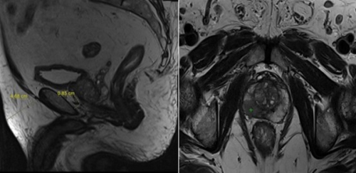
Objective : In this study, the relationship between periprostatic and subcutaneous fat thickness on MRI (magnetic resonans imaging) and Gleason scores (GS) was retrospectively evaluated. Materials and Methods: Fifty-one patients who underwent MRI before application of MRI fusion biyopsies were included 63,57±8,5 (45-88) years) in this study, On midsagittal T2- weighted MR images, the thickness of subcutaneous and periprostatic fat was calculated as the vertical distance from the pubic symphysis both to the skin and to the prostate, respectively. Results: The mean (±SD) subcutaneous fat thickness (SCFT) and periprostatic fat thickness (PPFT) were 17.38±13.02 mm and 5.64±3.89 mm, respectively. A positive correlation was found between Gleason scores and PPFT (p
Grand J Urol 2023;3(1):014-018, DOI: 10.5505/GJU.2022.52297
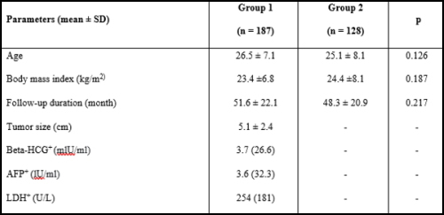
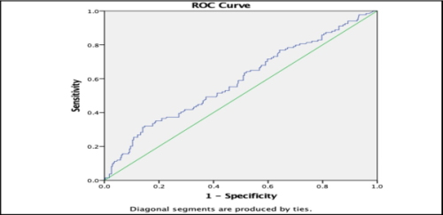
Objective: Recently, many hematological parameters have been frequently investigated as prognostic and predictive biomarkers for malignancies and inflammatory conditions, with the primary examples of these parameters being neutrophil-lymphocyte ratio (NLR), lymphocytemonocyte ratio (LMR), platelet-lymphocyte ratio (PLR), and mean platelet volume (MPV). In this study, we aimed to investigate the relationship between hematological parameters and prognosis in patients with a diagnosis of testicular malignancy (TM) by evaluating them in comparison with patients that underwent varicocelectomy as a control group. Materials and Methods: A total of 187 patients that underwent radical inguinal orchiectomy due to the diagnosis of testicular tumor were included in Group 1and 128 patients of similar age that underwent varicocelectomy using a similar incision in Group 2 as controls. Hematological and biochemical blood results were collected one day before radical orchiectomy. The parameters of NLR (neutrophil/ lymphocyte counts), PLR (neutrophil/ lymphocyte counts), PLR (platelet/ lymphocyte counts), and LMR (lymphocyte/ monocyte counts) were expressed as the ratios of indicated blood cell components. Following standard inguinal radical orchiectomy, T staging was performed based on the final histology samples, and clinical N and M staging using imaging modalities. Results: The mean (± SD) ages of the patients were 26.5±7.1, and 25.1±8.1 years for Groups 1 and 2, respectively. The neutrophil counts, NLR, and PLR were significantly higher in Group 1, while the lymphocyte count, MPV, and LMR were significantly higher in Group 2 (pT1 stage TM (pT1 in terms of MPV and PLR. Conclusion: We determined NLR and LMR as parameters that can be used in the pathological staging of cases with TM. We consider that these hematological parameters have an important place in predicting the prognosis in this patient group.
Grand J Urol 2023;3(1):026-030, DOI: 10.5505/GJU.2023.74745
Objective: In this study, we aimed to investigate the predictive value of neutrophil-lymphocyte ratio (NLR) in determining progression and recurrence in non-muscle invasive bladder tumors (NMIBC). Materials and Methods: The files of patients who underwent transurethral bladder tumor resection (TUR-BT) in our urology clinic between 2015 and 2020 were reviewed retrospectively. A total of 405 patients were included in the study. The patients were evaluated, and grouped in terms of disease progression, and recurrence observed during follow-up. Elevation of T stage of the disease, low grade tumor progressing to a high grade, and carcinoma in situ (CIS) negative cases advancing into CIS-positive stage were considered as evidence of disease progression. Results: Fifty female, and 355 male patients were included in the study. The mean age of the study population was 64.9 ± 12.75 years. Disease recurrence was detected in 134, and disease progression in 136 out of 405 patients. The mean NLR value of the patients with relapse was 2.45 ± 2.75 (p=0.009). The mean NLRs of patients with, and without progression were 1.94 ± 1.67, and 2.04 ± 2.3, respectively (p=0.645). Conclusion: High NLR value can predict recurrence in the follow-up of NMIBC patients. The preoperatively detected higher NLR value is a warning for the physician and draws attention to the need for more invasive and regular follow-up of the patient.
Grand J Urol 2024;4(2):035-039, DOI: 10.5505/GJU.2024.18189
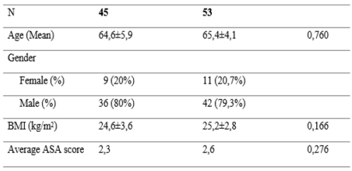
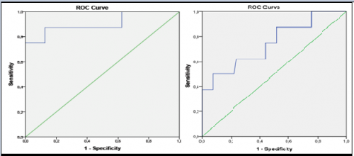
Objective: Radical cystoprostatectomy is the most important treatment option in local control and standard surgical treatment in muscle-invasive bladder cancer, and also has serious complications that occur during the postoperative period. We have aimed to evaluate postoperative bowel complications with peritoneal closure-assisted ileal conduit extraperitonealization technique. Materials and Methods: The data of 98 patients who underwent radical cystectomy and ileal conduit urinary diversion with the diagnosis of non-metastatic bladder cancer between 2015 and 2023 were retrospectively screened. The groups of patients who underwent extraperitonealization of the ileal conduit with radical cystectomy and traditional radical cystectomy were evaluated comparatively in terms of perioperative outcomes and postoperative complications. Results: Forty-five patients who underwent cystectomy with ileal conduit extraperitonealization technique and 53 patients who underwent ileal loop diversion with traditional cystectomy were evaluated comparatively. There was no statistically significant difference between the two groups in terms of demographic characteristics and duration of surgery. In the group that underwent cystectomy with extraperitonealization of the ileal conduit technique, the return of the normal gas pattern and the dwell time of the nasocracymic tube were statistically significantly shorter than the group that did not (p=0.017, p=0.023). The average length of hospital stay was 7.2 days in the extra-peritonealization group and 14.1 days in the group that did not undergo extraperitonealization, and this period was significantly shorter in the extraperitonealized group (p=0.013). There were no complications requiring reoperation in the extraperitonealized group. Conclusion: In radical cystectomy and ileal loop cutaneous urinary diversion, extraperitonizing the ileal segment reduces postoperative intestinal complications.
Grand J Urol 2021;1(2):43-48, DOI: 10.5222/GJU.2021.58066
Objective: Levels of insulin-like growth factor 1 (IGF-1) have been associated with prostate carcinoma. We have investigated whether IGF-1 level has an early predictive value for the biochemical relapse in the prostate carcinoma patients with a negative surgical margin who underwent curative surgery. Materials and Methods: We retrospectively analyzed 82 patients who were followed-up regularly and did not receive neoadjuvant or adjuvant chemotherapy. We classified patients as having Gleason scores ≥7 and
Grand J Urol 2022;2(2):047-052, DOI: 10.5505/GJU.2022.02986
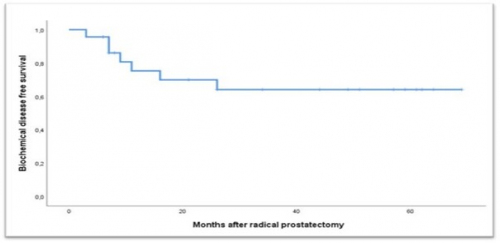
Objective: To evaluate the impact of radical prostatectomy (RP) and extended pelvic lymph node dissection (EPLND) on the disease process in terms of oncological outcomes and quality of life in the treatment of clinical stage C T3N0M0 prostate cancer (PCa). Materials and Methods: The data of patients with cT3N0M0 who had undergone open radical prostatectomy and extended pelvic lymph node dissection in our clinic between January 2015 and March 2021 were analyzed retrospectively. Preoperative and postoperative data were compared in terms of oncological and functional outcomes. Biochemical recurrence was accepted as detection of PSA >0.2 ng/ml on consecutive measurements and biochemical disease-free survival time was calculated. Results: The mean age of 23 operated patients who met the study criteria, was 66.8±7.4 years. In the pathological staging, the organ-confined disease was detected in 10 (43.4%) patients. Surgical margin positivity was observed in 6 (26.2%), while lymph node positivity in 3 (13.1%) patients. Biochemical recurrence was detected in 7 (30.2%) patients during a mean follow-up period of 33.6±22.9 months. The mean biochemical disease-free survival time was 48.4±6.3 months. In the evaluations of the patients at the postoperative 6th months, a 3.2±2.2-point decrease was found in the International Prostate Symptom Score (IPSS) (p=0.001) and a 13.1±5.0 point decrease in the International Index of Erectile Function (IIEF) score (p=
Grand J Urol 2021;1(2):66-70, DOI: 10.5222/GJU.2021.39974
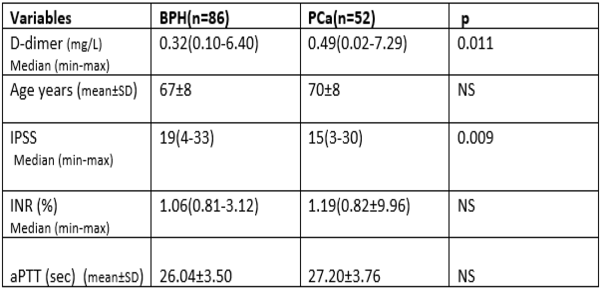
Objective: To investigate the value of D-dimer, a marker of fibrinolysis, in metastatic and non-metastatic prostate cancer. Materials and Methods: A retrospective analysis was performed on 138 male patients including 52 patients with prostate cancer and 86 with benign prostatic hyperplasia. Participants who had factors that altered D-dimer levels were excluded. The mean ages of the groups were similar (70 ± 8 vs 68 ± 8, p= NS). In addition, data regarding biochemical findings, prostate-specific antigen and hemostatic markers, including D-dimer, were retrieved from the database of our hospital. The cut-off point of D-dimer was 0.5 mg/L. Data from scintigraphy and magnetic resonance imaging (MRI) scans related to metastasis were also considered. Patients who showed findings of metastasis according to scintigraphy and lumbosacral MRI were accepted as having metastases. Positive findings in only scintigraphy in any area were considered suspicious for metastasis. Results: Patients with prostate cancer had higher D-dimer levels than benign prostate hyperplasia patients (p= 0.024). Sixteen patients with metastatic prostate cancer and suspicious for metastasis had markedly high D-dimer levels compared to benign prostate hyperplasia and non-metastatic prostate cancer patients. Conclusion: prostate cancer, especially when metastatic, may increase D-dimer levels.
Grand J Urol 2024;4(3):070-076, DOI: 10.5505/GJU.2024.18894
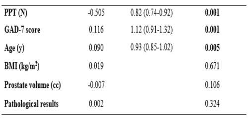
Objective: To evaluate whether the assessment of anxiety and pain perception before a biopsy procedure may predict patients" perceived pain scale scores during transrectal ultrasound-guided prostate biopsy. Materials and Methods: Patients who were administered the Mini-Mental State Examination 24 h before the biopsy were evaluated based on electrically and mechanically induced pain thresholds. Patients were assessed with Generalized Anxiety Disorder (GAD)-7 scale sscores in the hour before biopsy. The pain experienced by patients during rectal probing and biopsy was assessed using Visual Analog Scale (VAS) scores. Results: The mean age and median PSA level of the patients were 65.52 ± 7.85 years and 9.73 (1.4-155) ng/dL, respectively. The median VAS scores during rectal probing and biopsy were 3 (0-10) and 4 (0-10) respectively. VAS scores calculated during procedures were moderately-to-strongly correlated with the index finger of mechanically induced pain pressure threshold (PPT) (r=−0.606, p=0.001 and r=−0.760, p=0.001). Multiple regression analyses revealed that severity of the intraprocedural pain was correlated with age, GAD-7, and PPT index finger scores (p=0.005, p=0.001, p=0.001, respectively). A correlation was noted between the refusal of repeat prostate biopsy and higher pain scores (p
Grand J Urol 2024;4(3):083-088, DOI: 10.5505/GJU.2024.54227
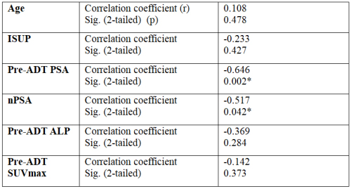
Objective: We sought to identify predictive factors affecting time to castration resistance in metastatic prostate cancer patients receiving androgen deprivation therapy (ADT). Materials and Methods: We retrospectively evaluated 47 patients who received ADT with the diagnosis of metastatic prostate cancer. The patients" age, International Society of Urological Pathology (ISUP) scores, baseline prostate specific antigen (PSA), alkaline phosphatase (ALP) value, prostate-specific membrane antigen (PSMA) tracer expression, represented by the maximum standardised uptake value (SUV max) at diagnosis, nadir PSA value, and time to resistance to ADT were recorded. Results: All patients included in the study were resistant to treatment with ADT. The mean age of the patients was 70.81 ± 1.15 years. The mean time to develop resistance to treatment after castration was 31.51 ± 4.9 months. In the correlation analysis, a significant negative correlation was detected between PSA, nadir PSA values and time to treatment resistance. The relationship between the SUVmax value of the primary prostate lesion, ALP value at the time of diagnosis and time to response to ADT developed was not significant. Conclusion: We found PSA values at diagnosis and nadir PSA values during follow-up to be predictive factors of treatment resistance in metastatic prostate cancer patients receiving ADT.
Grand J Urol 2022;2(3):100-107, DOI: 10.5505/GJU.2022.74755
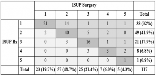
Objective: The primary aim of this study is the determination of International Society of Urological Pathology (ISUP) grade group (GG) upgrading prevalence and its risk factors in prostate cancer patients. Materials and Methods: This study was conducted on 117 patients who all underwent open radical prostatectomy in our institution between 2011 and 2020. Patients who received neoadjuvant therapy prior to surgery and had metastasis in lymph nodes or bones were excluded from the study. Results: In 28 (23.9%) cases ISUP GG had upgraded in final pathology. While grade group of 81 (69.2%) patients did not change, it was downgraded in the remaining 8 (6.8%) cases. In the univariate analysis for the predictors of ISUP GG upgrade, ISUP GG distribution in biopsy pathology (OR: 0.46, 95% CI: 0.26-0.82, p=0.009), positive core fraction (PCF) (OR: 0.07, 95% CI 0.01-0.85, p=0.037), greatest positive core percentage (GPC) (OR: 0.12, 95% CI: 0.02- 0.68, p=0.016) and extraprostatic invasion extended (EPI-extended) (OR: 2.95, 95% CI: 1.16- 7.49, p=0.023) were all identified as significant factors. When these significant factors were analyzed in multivariate logistic regression analysis, biopsy ISUP grade (OR: 0.38, 95% CI: 0.18-0.79, p=0.01), greatest percentage of cancer (GPC) (OR: 0.10, 95% CI 0.01-0.78, p=0.027) and EPI-extended (OR 14.9, 95% CI:3.1-71.9, p=0.01) were shown as independent predictors. Conclusion: ISUP GGs of a significant number of patients upgrade in the final pathology. Initial biopsy ISUP score and greatest positive core percentage in the biopsy are independent predictors of ISUP GG upgrade risk. EPI-extended was also significantly higher in ISUP upgrade group. Tumor upgrade risk should be considered prior to prostate cancer treatment.
Grand J Urol 2021;1(3):101-108, DOI: 10.5222/GJU.2021.40085
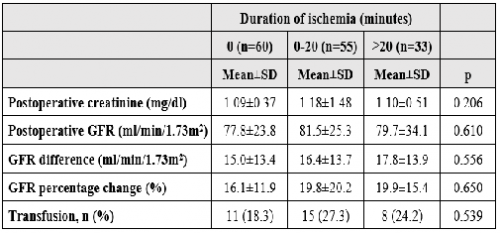
Objective: To specify the prognostic factors predicting complication rates and postoperative renal function in patients operated with partial nephrectomy. Materials and Methods: Our health center"s archive system was scanned retrospectively for the time interval between January 2006- January 2021 for patients operated with partial nephrectomy for renal mass. History, comorbidities and laboratory results, operational information, tumor morphologies in radiographic images and its specified scores (R.E.N.A.L. score, PADUA score, C-index), peroperative and postoperative complications and pathology results of 148 regularly followed-up patients were analyzed. Results: Mean age of the patients was 55.04±10.91 years, ratio of male to female was 1.27 and mean tumor size was 3.56 cm. Mean follow-up period was 55.53±42.26 months. Postoperative creatinine value in the 6th month showed an increase of 0.18 mg/dl compared to preoperative value. Estimated glomerular filtration rate (eGFR) also decreased by an average of 18.3%. Operation of grade 4 tumors significantly affected the postoperative renal function. PADUA score (p=0.023) had a significant effect on postoperative GFRs and duration of ischemia. Also, difference in pre-and postoperative GFRs and its percentage change were significantly affected by C-index (p=0.035, p=0.042). Pathological size (p=0.038), R.E.N.A.L. score (p=0.001), PADUA score (p
Grand J Urol 2023;3(3):109-112, DOI: 10.5505/GJU.2023.35229
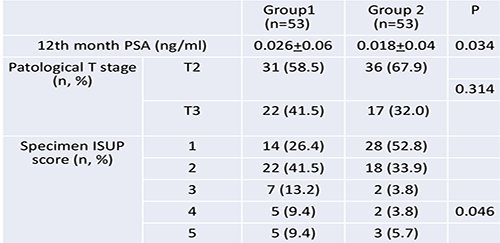
Objective: To examine the relationship between prostate weight and oncological and functional outcomes of Retzius-sparing robot-assisted radical prostatectomy (RS-RARP). Materials and Methods: Data of the patients who underwent RS-RARP in our clinic between December 2018 and December 2020 were evaluated retrospectively. A total of 106 patients with 12-month postoperative follow-up data were included in the study. The patients were separated into 2 groups according to the weights of the pathology specimens as Group 1 (n=53, prostate weight less than 50 g), and Group 2 (n=53, prostate weight more than 50 g). Postoperative oncological and functional data were analyzed. At the end of the 12th month, continence was regarded as requirement of no pad or 1 pad per day. Potency was considered as the ability to have sexual intercourse. Prostate-specific antigen (PSA) above 0.2 ng/ml in the follow-up period was considered as biochemical recurrence. Results: Preoperative PSA levels were comparable between groups (9.78+7.84 ng/ml vs. 11.87+8.38 ng/ml). There was no difference in clinical cancer stages and The International Society of Urological Pathology (ISUP) scores between the groups. Median vesicourethral anastomosis time (30 minvs.33 min) and median operative time (240 min vs. 240 min) were comparable in both groups (p>0.05). There was no difference in localized disease and locally advanced disease rates between the groups (pT2: 58.5% vs. 67.9%, pT3: 41.5% vs. 32.0%). Respective surgical margin positivity (SMP) ([16.9% (n=9) vs 9.4% (n=5]), and 12th month biochemical recurrence rates (11.32% vs 3.77%) for Groups 1 and Group 2, were as indicated (p>0.05). Postoperative urinary continence rates at 12 months were 89% and 90% in Groups 1 and 2, respectively (p>0.05). Continence status was not different between the groups. Potency rates at 12 months were comparable between the groups. Conclusion: RS-RARP can be applied in patients with any size of prostates with comparable functional and oncological outcomes.
Review Article
Prostate cancer is one of the most common tumor in males. Radical prostatectomy, radiotherapy and watchful waiting are the main treatment options in localized disease. Radiotherapy together with hormonotherapy is accepted as the standard of care in patients with advanced stages. Surgery or radiotherapy has comparable local control and survival outcomes in localized disease. During recent years a significant reduction in the rate of serious side effects has been achieved due to the development of modern radiotherapy techniques. With the use of these techniques such as Intensity-modulated radiotherapy (IMRT), Image-guided radiotherapy (IGRT), Stereotactic body radiotherapy (SBRT), high doses can be given safely and the rates of serious short-or long-term side effects have not exceeded 1 percent. Modern radiotherapy techniques allow dose escalation for the target volume, and due to its achievement of sharp dose gradient around the target volume and enable to increase radiation doses homogeneously within the target volume without exceeding the tolerance doses in organs at risk. In the last few years hypofractionation has gained popularity in the curative radiotherapy of prostate cancer.
Focal ablation therapies in prostate cancer have been actively evaluated in the light of recent literature. According to published data,focal ablation therapies appear to be well tolerated and have an acceptable side effect profile. Moreover, while clinical outcomes were not homogenous, short-term oncological results of some focal ablation therapies such as laser and irreversible electroporation (IRE) have been found as good as curative ones. While waiting long-term oncological results, focal ablation therapies in prostate cancer are beinmg used increasingly.
Case Report
Grand J Urol 2024;0(0):, DOI: 10.5505/GJU.2025.24008
Upper urinary tract urothelial tumors are rarely seen. Only 5% of all transitional cell carcinomas (TCCs) originate from the upper urinary tract. Although skin metastases in urinary tumors are quite rare, the incidence of skin metastases in upper urinary tract TCCs is not exactly known due to the lack of published research on this subject. In this article, we aimed to present a lesion consistent with TCC metastasis in the left inguinal region of a patient who was operated on for a renal pelvic mass. A 61-year-old male patient underwent left laparoscopic nephrectomy because of a mass reported as renal cell carcinoma (RCC) in the upper pole of the left kidney. After the pathology result reported the presence of left renal TCC, the patient underwent left ureterectomy plus cuff resection in an another medical center. After this second operation, the skin lesion developing at the trocar entry site on the left inguinal region of the patient was reported as an abscess on imaging. The histopathology of the excised abscess was reported as TCC metastasis. There is no consensus on the treatment regimen for skin metastases of urothelial carcinomas, and these metastases have a relatively poor prognosis. One -year survival rate is generally 35%. The patient in our case received gemcitabine and cisplatin treatment and has been under oncology follow-up for twelve months.
Nephroblastoma (Wilms Tumor / WT) is the rare but the most common primary kidney tumor in children. The WT is generally diagnosed between 1 and 5 and the most common diagnosis is the age of 3. Up to 95% of WTs are diagnosed correctly with visualization (ultrasound, computed tomography and magnetic resonance). Histopathologicaly tissue examination is implemented and subtypes are determined. Surgery is one of the key factors in WT treatment. Transperitoneal radical nephrectomy is the standard operation for unilateral WTs. Nephron sparing surgery (partial nephrectomy) is suggested only in selected cases with a single kidney or bilateral WT. Other treatment combination for Wilms tumor involves chemotherapy and radiation treatment. A patient who was diagnosed with Wilms tumor in the right kidney and underwent partial nephrectomy which is rarely implemented or recommended in selected cases is represented in this article.
Chest wall malignancies are rare, constituting only 1% of all cancers. Prostate cancer, the second most common in men, typically metastasizes to bones, lymph nodes, and organs. However, sternum involvement is exceptionally rare, particularly with osteolytic metastasis. We report a 75-year-old man, clinically presenting with large anterior chest wall mass, which on further investigation revealed an expansile osteolytic sternal body metastasis from prostate cancer. This rarity poses diagnostic and therapeutic challenges, as documented cases of osteolytic secondaries to the sternum are scarce in medical literature. Diagnostic efforts involved comprehensive imaging and biopsy, confirming prostate cancer metastasis. Management requires a multidisciplinary approach, balancing effective cancer control with preserving the patient"s quality of life through systemic therapies, radiation, and surgery. The scarcity of osteolytic sternal body metastasis in reported cases highlights the atypical pattern of metastasis in prostate cancer emphasizing the need for a deeper understanding and contributing to the knowledge of this uncommon manifestation.
Grand J Urol 2024;4(1):032-034, DOI: 10.5505/GJU.2024.73745
Amyloidosis is related to the extracellular deposition of abnormal protein fibrils in various tissues. It is clinically interesting that such cases" clinical, radiological, and even endoscopic presentation mimic urothelial carcinoma to a great extent. Here, we discuss a case of a 34-year-old gentleman who presented with frank painless hematuria. The patient was diagnosed with a bladder mass suspicious of malignancy depending on the clinical presentation aided by the cystoscopic and radiological evaluation. Histopathologic samples of the transurethral resection of the mass proved to be primary bladder amyloidosis. Further investigations of systemic illness excluded the secondary amyloidosis. The purpose of this case presentation is to create awareness among the urologists to think for the rare entity of urinary amyloidosis especially if the histopathology is negative for the malignant cells.
Grand J Urol 2021;1(1):33-36, DOI: 10.5222/GJU.2021.98609
Retroperitoneal liposarcoma (RPLS) is a rare tumor. Early diagnosis and treatment are difficult due to absence of specific clinical presentations. We report a case of a 66-years-old woman who succesfully underwent complete surgical resection for a giant retroperitoneal liposarcoma. The complete surgical resection is the most important predictor of local recurrence and overall survival. We believe that complete surgical resection involving adjacent organs is a curative treatment to increase overall survival, especially in the presence of invasion of large tumors.
Non-germ cell testicular tumors are rarely seen. Sex-cord stromal tumors, which make up the majority of testicular tumors other than germ cell tumors, share common immunohistochemical and histomorphological features. Our aim in presenting this case is to define the Sertoli cell nodule (SCN), which is one of the relatively rare primary testicular pathologies that can be detected in half of adult undescended testes, with its histomorphological and immunohistochemical features, and to make its differential diagnosis.
Grand J Urol 2022;2(1):38-41, DOI: 10.5505/GJU.2022.70299
Immunosuppressive therapy is related to the increasing frequency of malignancies after transplantation. A small percentage (4.6%) of malignancies seen in kidney transplant patients are renal cell carcinomas (RCC) which occur almost exclusively in native kidneys. The prognosis of RCC largely depends on the presence of metastasis. Metastatic disease is very rare in small renal masses. In this case report, we aimed to present our case of approximately 4 cm-mass of metastatic RCC in our kidney transplant patient. During the examination due to exhaustion and weight loss, multiple suspicious metastatic lesions were observed in non-contrast computed tomography. In the patient who had multiple bone metastases on the whole-body bone scintigraphy, prostate cancer metastasis was considered in the first plan due to a history of prostate cancer before transplantation. This diagnosis could not be supported with prostate-specific membrane antigen-positron emission tomography/computed tomography (PSMA-PET/CT) scan. Whole body 18F-fluorodeoxyglucose positron emission tomography/computed tomography (18F-FDG PET/CT) was performed. A hypermetabolic mass lesion in the left kidney, multiple hypermetabolic lesions in the liver, in the left aortorenal junction, and in the skeletal system were observed. A biopsy was performed from the metastatic mass in the right lobe of the liver and the result was reported as renal cell carcinoma metastasis. Immunohistochemistry evaluation demonstrated positive staining for PAX-8, CK19, CD10 and negative staining for CK7, CK20, GATA-3, NAPSIN A, TTF-1, PSAP, glutamine synthetase and arginase. With all these findings, it was thought that the primary of metastases was the 4-cm mass in the left native kidney.
Angiomyolipomas are the most common benign mesenchymal tumors of the kidney. Although they are often seen sporadically, they can also be observed as a part of the tuberous sclerosis complex (TSC). They occur at an earlier age in cases associated with tuberous sclerosis (TS), bilateral mass and epithelioid formation. There are various treatment approaches such as active surveillance, nephron-sparing surgery, nephrectomy, angioembolization, and use of mammalian target of rapamycin (mTor) inhibitors. Our case was a patient with bilateral multiple renal angiomyolipomas associated with TS. We applied mTOR inhibitor and angioembolization therapy to this patient. In our article, we tried to evaluate our success rate in our treatment and the treatment regimens to be applied in these patients.
Grand J Urol 2022;2(2):072-075, DOI: 10.5505/GJU.2022.18209
Cystic nephroma is a rare, multicystic, non-hereditary benign lesion that does not contain solid components. The fact that this entity can not be easily distinguished from other cystic tumors of kidney creates difficulties in diagnosis and treatment. We present a 37-year-old male case with incidentally detected two cystic masses in his left kidney. After nephron-sparing nephrectomy, the histopathology of one of the cystic mass of the patient was reported as renal cell cancer and the other as cystic nephroma. Cystic nephroma is a rare tumor of the kidney. Definitive diagnosis can be made histopathologically rather than using physical examination and imaging methods. We reviewed the literature by presenting our case that had 2 cystic lesions in the same kidney, one of which was renal cell cancer and the other was cystic nephroma.
Splenogonadal fusion (SGF) is a very rarely seen congenital anomaly localized usually in the left testis and mimics a testicular tumor. There are two subtypes of SGF, as continuous and discontinuous SGF. Continuous SGF can usually be detected in childhood. The less common discontinious SGF may not be detected until adulthood, and may be mistaken for testicular tumor and cause unnecessary orchiectomies. In this case report, we aimed to present a patient who underwent orchiectomy due to a left testicular mass associated with hydrocele and was found to have discontinuous SGF in his histopathological evaluation.
COVID-19 is a highly contagious disease. This condition affects the decision of both the patient and the surgeon about the surgery of newly diagnosed cancer patients and it may also result in delays in cancer surgeries because of the limitations in healthcare applications. In our particular case, it was aimed to present the transition of the cancer from the localized stage to distant spread stage cancer since the patient who was pre-diagnosed with RCC and who was recommended surgery, did not want to undergo surgery due to COVID-19 pandemic and its risks. Our case was a 49-year-old female patient. In her computed tomography, a 58x70 mm heterogeneously enhancing solid lesion which showed exophytic extension from the middle zone of the right kidney to the lower pole was observed. Surgery was recommended for the patient but the patient claimed that she did not want to undergo surgery due to the risk of COVID-19 pandemic. The patient made an application for the surgery 8 months later. The new magnetic resonance imaging of the patient showed that there was a mass lesion of approximately 76x76x80 mm in size, which involved middle-lower part of the right kidney and extended into the opening of the renal vein VCI by invading the renal vein. Radical nephrectomy and thrombectomy procedure was applied to the patient with RCC? tumor. Due to psychosocial problems caused by the pandemic, surgeries are delayed and an acceleration of the cancer progression from the localized stage to the distant spread stage occurs indispensably.
Grand J Urol 2024;4(3):098-102, DOI: 10.5505/GJU.2024.52714
Six percent of cases with renal cell carcinoma (RCC) can present with thrombus, and also invasion to renal vein, and atrium may be observed in 44% and 1-4 % of these cases, respectively. These cases require multidisciplinary management and surgery should be the first treatment option. However, if a tumor is considered unresectable or metastatic, systemic therapy can be considered in the first instance. Herein, we present 2 cases. A 77-year-old female patient presented with right renal tumor 89 mm in diameter with thrombus level IV considerably unresectable started to receive treatment with nivolumab and cabozantinib. After 6 months of treatment thrombus was reduced to level II. A 43-year-old male, presented with 110 mm- right renal mass with thrombus level II and lung metastases. He started to receive pembrolizumab and axitinib. At 6 months of treatment, the size of the tumor and thrombus decreased. In both cases we performed laparoscopic radical nephrectomy with thrombectomy, and pathology reports indicated the presence of clear cell RCC, Grade 3, pT3b-Nx. Systemic treatment in patients with RCC associated with tumor thrombus, whether metastatic or not, would seem to obtain some benefit prior to surgery -line favor surgical feasibility.
Extra-uterine leiomyomas of urethral origin are rarely encountered neoplasms possessing unique features such as the characteristic growth pattern, diagnostic challenges owing to a long list of possible differential diagnoses, possible cure with surgical management and the unique complications that accompany surgical management. Herein, we report a case of urethral leiomyoma in a middle- aged woman with a brief discussion on the evaluation and management aspects including a concise description of this pathology based on the scarce literature information available.
Grand J Urol 2022;2(3):117-121, DOI: 10.5505/GJU.2022.58068
Paratesticular liposarcomas (PLSs) are mostly painless, slow-growing and extremely rare inguinal or scrotal masses. Reports of approximately 270 cases have been published in the literature so far, but only a few of them contain information about giant PLSs exceeding 10 cm in size. Correct diagnosis and treatment is important as PLSs tend to cause local relapses and distant metastases. Here, we aimed to present, and evaluate a dedifferentiated (24 cm), and a well-differentiated (12 cm) giant PLS in the light of the literature data.
Grand J Urol 2021;1(3):142-145, DOI: 10.5222/GJU.2021.22931
Primary or secondary lymphoma of the prostate is a rare condition. Mantle cell lymphoma (MCL) represent 4-9% of all lymphomas. Prostate involvement with MCL is very rare, with only 11 reported cases up to now. Here we present a case with lower urinary tract symptoms and prostate-specific antigen (PSA) elevation diagnosed with MCL of the prostate. Prostate biopsy was performed in a 70-year-old patient due to increased PSA. After the pathology result was reported as prostatic MCL, imaging studies and sampling of additional pathological specimens were performed for staging. An improvement was observed in the urinary system complaints of the patient who started chemotherapy regimen. While prostatectomy was performed in some of the prostatic MCL cases reported previously, in some, no additional treatment was required after chemotherapy. Our case is the only prostatic MCL case with elevated PSA levels, but did not receive the diagnosis of prostate cancer. Physicians should keep in mind that, prostatic MCL can present with nonspecific symptoms. Staging should be performed in patients whose histopathologic diagnosis is lymphoma of the prostate so as to determine appropriate treatment options.